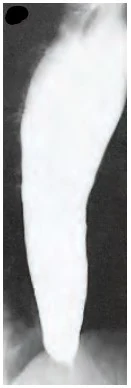
Esophagus

Oesophagus: Anatomy, Physiology, Diseases, and Surgical Insights

Anatomy and Physiology of the Oesophagus

The oesophagus is a muscular tube approximately 25-30 cm long in adults, connecting the throat (pharynx) to the stomach. It plays a crucial role in transporting food and liquids to the stomach through coordinated muscular contractions known as peristalsis. The oesophagus is made up of several layers:

  • Mucosa: The innermost lining that produces mucus to ease the passage of food.
  • Submucosa: A layer containing blood vessels, nerves, and glands.
  • Muscularis: Muscle layers responsible for peristalsis.
  • Adventitia: The outermost layer providing structural support.

Key physiological functions include the prevention of food reflux through the lower oesophageal sphincter and the protection of the mucosa from gastric acid.

Importance of the Oesophagus

The oesophagus is vital for digestion as it ensures the seamless passage of ingested food from the mouth to the stomach. Proper oesophageal functioning prevents regurgitation and aspiration, which can impact respiratory health.

Diseases of the Oesophagus

Common Conditions:

  1. Gastroesophageal Reflux Disease (GERD): Chronic acid reflux causing heartburn and damage to the oesophageal lining.
  2. Oesophagitis: Inflammation of the oesophagus due to infections, medications, or acid reflux.
  3. Barrett's Oesophagus: A condition where the oesophageal lining changes, increasing cancer risk.
  4. Oesophageal Strictures: Narrowing of the oesophagus due to scarring.
  5. Oesophageal Varices: Enlarged veins in the lower oesophagus, often due to liver disease.
  6. Oesophageal Cancer: Can be squamous cell carcinoma or adenocarcinoma.
  7. Achalasia: A rare disorder where the oesophageal muscles fail to relax, causing difficulty swallowing.


Esophageal varices

Achalasia of esophagus

Barrett's esophagus

Diseases Requiring Surgery and Surgical Indications

  1. GERD: Surgery may be needed if medications fail or complications like strictures develop.
  2. Oesophageal Cancer: Surgery is often necessary to remove cancerous tissues.
  3. Achalasia: Surgical intervention is required when other treatments are ineffective.
  4. Barrett's Oesophagus: In severe cases with dysplasia, surgery is recommended.
  5. Oesophageal Perforations: Emergency surgery is required to repair tears.

Overview of Common Surgeries

  1. Fundoplication: Wrapping the upper part of the stomach around the oesophagus to prevent reflux.
  2. Oesophagectomy: Removal of the oesophagus, often for cancer treatment.
  3. Heller Myotomy: Cutting the muscles at the lower oesophageal sphincter to treat achalasia.
  4. Endoscopic Procedures: Minimally invasive techniques for Barrett’s oesophagus and small tumours.
  5. Stent Placement: Used to alleviate obstructions.

Pre-Surgery Preparation

  • Medical Evaluations: Blood tests, imaging studies, and endoscopy.
  • Dietary Modifications: Liquid or low-residue diet before surgery.
  • Medication Adjustments: Stopping certain medications that increase bleeding risk.
  • Smoking Cessation: Essential for better surgical outcomes.
  • Counseling: Psychological support for major surgeries.

Risks and Complications of Each Surgery

  1. Fundoplication: Difficulty swallowing, gas bloat syndrome.
  2. Oesophagectomy: Risk of infection, anastomotic leakage, breathing difficulties.
  3. Heller Myotomy: Reflux as a common side effect.
  4. Endoscopic Procedures: Bleeding, perforation.
  5. Stent Placement: Migration of the stent, infection.

Recovery Process for Each Type of Surgery

  1. Fundoplication: Hospital stay of 1-3 days; soft diet for a few weeks.
  2. Oesophagectomy: Prolonged recovery with several weeks in the hospital and dietary adjustments.
  3. Heller Myotomy: Recovery within a week; gradual dietary changes.
  4. Endoscopic Procedures: Short recovery period; same-day discharge in most cases.
  5. Stent Placement: Minimal recovery time; dietary adjustments.

Success Rates and Benefits of Each Surgery

  1. Fundoplication: Success rate over 90% for GERD relief.
  2. Oesophagectomy: Improved survival rates in early-stage cancer.
  3. Heller Myotomy: 85-95% success in relieving achalasia symptoms.
  4. Endoscopic Procedures: High success for early cancer and Barrett’s oesophagus.
  5. Stent Placement: Effective for immediate symptom relief.

Latest Innovations and Advancements

  • Robotic Surgery: Improved precision and faster recovery.
  • Endoscopic Therapies: Radiofrequency ablation for Barrett’s oesophagus.
  • Minimally Invasive Techniques: Reduced complications and hospital stays.
  • 3D Imaging and Navigation: Enhanced surgical accuracy.

Expert Opinions for Each Surgery

  1. Fundoplication: "It's a reliable solution for patients who don't respond to medications," says Dr. John Smith, Gastroenterologist.
  2. Oesophagectomy: "Early detection and surgical intervention offer the best outcomes," notes Dr. Sarah Lee, Oncologist.
  3. Heller Myotomy: "Patients experience significant symptom relief," emphasizes Dr. James White, Surgeon.
  4. Endoscopic Procedures: "Minimally invasive techniques are game-changers for early-stage diseases," highlights Dr. Maria Gonzalez, Endoscopist.

Facts to Remember

  • The oesophagus plays a crucial role in digestion.
  • Early detection of oesophageal diseases can significantly improve outcomes.
  • Minimally invasive surgeries have shorter recovery times.

Frequently Asked Questions (FAQs)

  1. Can GERD be treated without surgery? Yes, through medications and lifestyle changes.
  2. What is the recovery time for oesophagectomy? It can take several months to fully recover.
  3. Are oesophageal surgeries painful? Pain management strategies are used to minimize discomfort.

Legal and Ethical Considerations

  • Informed consent is essential before any surgical procedure.
  • Patients have the right to seek second opinions.
  • Surgeons must adhere to ethical guidelines and safety protocols.

Summary

The oesophagus is a critical component of the digestive system, and various diseases can affect its function. While many conditions are treatable with medications and lifestyle changes, surgery is sometimes necessary. Advances in surgical techniques and technology have significantly improved outcomes and recovery times. Understanding the anatomy, diseases, and treatment options empowers patients to make informed decisions.

References

  1. Smith, J., & Lee, S. (2022). Gastrointestinal Surgery: Comprehensive Guide. Medical Publishers.
  2. American Gastroenterological Association (2023). Oesophageal Health and Diseases. Retrieved from www.gastro.org
  3. National Cancer Institute (2023). Oesophageal Cancer Treatment Guidelines. Retrieved from www.cancer.gov
  4. Gonzalez, M. (2021). "Minimally Invasive Endoscopic Techniques," Journal of Gastroenterology.
  5. World Health Organization (2023). Global Guidelines for Digestive Health.

Lignocaine

Lignocaine (Lidocaine)

Lignocaine (Lidocaine) is a widely used amide local anesthetic and Class IB antiarrhythmic agent. It stabilizes neuronal membranes by inhibiting sodium ion influx, which prevents nerve signal transmission. As an antiarrhythmic, it reduces cardiac excitability and electrical conduction in ischemic tissues.

Uses

  • Local Anesthetic: Used for local and regional anesthesia in minor surgeries, dental procedures, and nerve blocks.
  • Antiarrhythmic: Effective in the management of ventricular tachycardia and ventricular fibrillation.
  • Adjunct in Intubation: Reduces intracranial pressure during tracheal intubation.
  • Pain Management: Utilized for postoperative and chronic pain relief.

Dosage and Administration

Local Anesthesia (Plain)

  • Adults: 2-3 mg/kg (maximum dose: 300 mg).
  • Pediatrics: 1-1.5 mg/kg.

Local Anesthesia (With Epinephrine)

  • Adults: 5-7 mg/kg (maximum dose: 500 mg).
  • Pediatrics: 3-4 mg/kg.

Antiarrhythmic Use

  • Adults (IV): 1-1.5 mg/kg IV bolus. Repeat doses of 0.5-0.75 mg/kg every 5-10 minutes if needed (maximum 3 mg/kg).
  • Pediatrics: 1 mg/kg IV bolus followed by infusion at 20-50 mcg/kg/min.

Toxic Dose

  • Plain: >3 mg/kg.
  • With Epinephrine: >7 mg/kg.
  • Toxic Plasma Concentration: >5 mcg/mL.

Dose Adjustment in Different Diseases

  • Hepatic Impairment: Reduced clearance; lower doses recommended.
  • Renal Impairment: Minimal impact, but accumulation may occur with long-term use.
  • Heart Failure: Use with caution due to decreased hepatic perfusion.

Presentation or Form

  • Injectables: 0.5%, 1%, and 2% solutions in 5 mL and 10 mL ampoules.
  • Topical: 5% lidocaine patches, EMLA cream (2.5% lidocaine + 2.5% prilocaine).
  • Other Forms: Lidocaine gels, sprays, and ointments.

Pharmacokinetics

  • Absorption: Rapid absorption when administered parenterally.
  • Distribution: Widely distributed; 60-80% plasma protein binding.
  • Metabolism: Primarily in the liver by CYP1A2 and CYP3A4 enzymes.
  • Excretion: Renal; 90% excreted as metabolites.
  • Half-life: 90-120 minutes.

Pharmacodynamics

  • Mechanism of Action: Inhibits voltage-gated sodium channels, blocking nerve signal transmission. In cardiac tissue, it reduces excitability and conduction, particularly in ischemic areas.
  • Onset: 2-5 minutes.
  • Duration: 30 minutes to 3 hours (longer with epinephrine).

Drug Interactions

  • CYP3A4 Inhibitors (e.g., ketoconazole): Increase the risk of toxicity.
  • Phenytoin: Decreases lidocaine efficacy by enhancing metabolism.
  • Beta-blockers: May reduce lidocaine clearance.
  • Other Local Anesthetics: Increased risk of systemic toxicity.

Comparison with Other Drugs in the Same Category

  • Bupivacaine: Longer duration but higher cardiotoxicity.
  • Ropivacaine: Similar duration with a better safety profile for cardiac toxicity.
  • Mepivacaine: Faster onset but shorter duration compared to lidocaine.

Precautions and Special Considerations

  • Allergic Reactions: Avoid in patients with hypersensitivity to amide-type local anesthetics.
  • Cardiac Conditions: Use cautiously in heart block and bradycardia.
  • Pregnancy: Category B; safe when used appropriately.
  • Elderly: Lower doses recommended due to reduced hepatic metabolism.
  • Liver Disease: Dose adjustments are necessary.

Side Effects

  • Common: Dizziness, drowsiness, nausea, paresthesia.
  • Serious: Seizures, hypotension, bradycardia, arrhythmias.
  • Local Anesthesia Systemic Toxicity (LAST):
    • Early Symptoms: Tinnitus, circumoral numbness, metallic taste.
    • Advanced Symptoms: Seizures, coma, bradycardia, arrhythmias.
    • Management: Lipid rescue therapy (20% lipid emulsion)
      • Bolus: 1.5 mL/kg over 1 minute.
      • Infusion: 0.25 mL/kg/min for 30-60 minutes.

Recent Updates and Guidelines

  • American Heart Association (AHA) Guidelines: Lidocaine is recommended as an alternative to amiodarone for refractory ventricular fibrillation or pulseless ventricular tachycardia.
  • Enhanced Formulations: Development of extended-release lidocaine patches for chronic pain management.
  • FDA Updates: Emphasis on appropriate dosing to prevent systemic toxicity.

Facts to Remember

  • Maximum safe dose: 3 mg/kg (plain) and 7 mg/kg (with epinephrine).
  • Onset within 2-5 minutes; duration up to 3 hours.
  • Effective in managing ischemia-induced ventricular arrhythmias.
  • Lipid rescue therapy is crucial for LAST management.

References

  1. Stoelting’s Pharmacology and Physiology in Anesthetic Practice, 5th Edition.
  2. NCBI StatPearls - Lidocaine: https://www.ncbi.nlm.nih.gov/books/NBK539881/
  3. Local Anesthetic Systemic Toxicity - BJA Education: https://bjaed.org/article/S2058-5349(19)30040-0/fulltext
  4. American Heart Association Guidelines for Advanced Cardiac Life Support.
  5. FDA Drug Safety Communications on Lidocaine Formulations.

Adenosine

Adenosine

Adenosine is a naturally occurring purine nucleoside with a pivotal role in cellular energy transfer and signal transduction. Clinically, it is widely utilized as an antiarrhythmic agent for the acute termination of paroxysmal supraventricular tachycardia (PSVT) and as a diagnostic tool in cardiology.

Uses

  • Supraventricular Tachycardia (SVT): Termination of paroxysmal SVT, including atrioventricular nodal reentrant tachycardia (AVNRT) and Wolff-Parkinson-White (WPW) syndrome.
  • Diagnostic Aid: Assists in revealing atrial activity (e.g., flutter, fibrillation) and differentiating SVT from ventricular arrhythmias.
  • Wide-Complex Tachycardia: Used as a diagnostic tool to differentiate ventricular tachycardia (VT) from SVT with aberrancy.
  • Stress Testing: Pharmacologic stress agent for myocardial perfusion imaging.

Dosage and Administration

Adults:

  • SVT:
    • Initial: 6 mg rapid IV bolus followed by a 20 mL saline flush.
    • If unresponsive, a second dose of 12 mg can be administered after 1-2 minutes.
    • If needed, a third 12 mg dose may be given.
  • Cardiac Stress Testing:
    • Infusion: 140 mcg/kg/min IV over 4 to 6 minutes.

Pediatrics:

  • SVT:
    • Initial dose: 0.1 mg/kg IV bolus (maximum 6 mg), followed by a saline flush.
    • Second dose: 0.2 mg/kg IV (maximum 12 mg) if necessary.
    • Maximum dose: 12 mg.

Administration Notes:

  • Administer via a large-bore IV in a proximal vein (e.g., antecubital fossa).
  • Always follow with a rapid saline flush.
  • Continuous ECG monitoring during and after administration is essential.

Dose Adjustment in Different Conditions

  • Renal Impairment: No dose adjustment required.
  • Hepatic Impairment: No dose adjustment required.
  • Heart Transplant Recipients: Lower doses may be needed due to heightened sensitivity.

Presentation and Form

  • IV Solution: 3 mg/mL in 2 mL and 4 mL vials.

Pharmacokinetics

  • Absorption: Administered intravenously, achieving immediate effects.
  • Onset: 10 to 20 seconds.
  • Duration: Less than 1 minute.
  • Metabolism: Rapidly metabolized by adenosine deaminase in erythrocytes and vascular endothelial cells.
  • Excretion: Metabolites are excreted in the urine.

Pharmacodynamics

  • Adenosine binds to A1 receptors in the AV node, causing transient AV block and interruption of reentrant circuits.
  • Additionally, it activates A2 receptors, leading to coronary vasodilation.

Drug Interactions

  • Dipyridamole: Potentiates adenosine—consider dose reduction.
  • Theophylline and Caffeine: Antagonize adenosine—may require higher doses.
  • Carbamazepine: Increases risk of heart block.
  • Beta-Blockers and Calcium Channel Blockers: May exacerbate bradycardia.

Comparison with Other Drugs in the Same Category

  • Adenosine vs. Verapamil: Adenosine has a faster onset and shorter duration, making it preferable for acute termination of SVT.
  • Adenosine vs. Amiodarone: Adenosine is more effective for AVNRT but less useful for atrial tachycardias.
  • Adenosine vs. Digoxin: Digoxin has a slower onset and is used for chronic rate control rather than acute arrhythmia termination.

Precautions and Special Considerations

  • Contraindications:
    • Second- or third-degree AV block (without pacemaker)
    • Sick sinus syndrome (without pacemaker)
    • Asthma or severe COPD (risk of bronchospasm)
    • Hypersensitivity to adenosine
  • Special Populations:
    • Pediatrics: Saline flush critical for effectiveness.
    • Transplanted Hearts: Lower doses may be necessary.
  • Monitoring: Continuous ECG monitoring is essential during administration.
  • Administration: Use a proximal IV site for rapid delivery.
  • Stress Testing: Administered as a continuous infusion rather than a bolus.

Side Effects

Common:

  • Flushing
  • Chest pain
  • Shortness of breath (SOB)
  • Dizziness

Serious:

  • Bronchospasm (especially in patients with asthma)
  • Bradycardia
  • Hypotension
  • Transient asystole

Rare:

  • Proarrhythmic events, including atrial fibrillation (especially in WPW syndrome)

Recent Updates and Guidelines

  • 2023 ACC/AHA Guidelines: Adenosine remains the first-line treatment for acute termination of PSVT.
  • New Recommendations: Enhanced focus on ECG monitoring during administration to detect rare atrial fibrillation post-adenosine administration.

Key Facts to Remember

  • Ultra-Short Half-Life: Less than 10 seconds.
  • Selective AV Nodal Effect: Effective for terminating AVNRT and AVRT.
  • Rapid Onset: Effects are seen within 10 to 20 seconds.
  • ECG Monitoring: Essential during administration.
  • Diagnostic Utility: Helps unmask atrial arrhythmias and differentiate tachycardias.

References

  1. Adenosine - NCBI StatPearls https://www.ncbi.nlm.nih.gov/books/NBK519049/
  2. NCBI - Pharmacological Stress Testing https://www.ncbi.nlm.nih.gov/books/NBK555963/
  3. Goodman & Gilman’s The Pharmacological Basis of Therapeutics, 13th edition.
  4. Harrison’s Principles of Internal Medicine, 20th edition.
  5. PALS: Pediatric dosing guidelines for SVT.

Amiodarone

Amiodarone

Amiodarone is a broad-spectrum antiarrhythmic medication belonging to Class III of the Vaughan Williams classification. It is widely used to manage and treat various types of cardiac arrhythmias due to its unique pharmacological properties that also span Class I, II, and IV actions.

Uses:

  • Ventricular Arrhythmias: Effective in the treatment of ventricular tachycardia (VT) and ventricular fibrillation (VF).
  • Atrial Fibrillation (AF): Management of rhythm control in persistent and paroxysmal atrial fibrillation.
  • Cardiac Arrest: Part of advanced cardiac life support (ACLS) for shock-refractory VF or pulseless VT.
  • Supraventricular Tachycardia (SVT): Used for rate control and rhythm conversion.

Dosage and Administration:

Ventricular Fibrillation/Pulseless VT (ACLS)

  • IV Bolus: 300 mg IV push, followed by a second dose of 150 mg if necessary.

Ventricular or Supraventricular Arrhythmias:

  • IV Loading Dose: 150 mg IV over 10 minutes, followed by 1 mg/min infusion for 6 hours, then 0.5 mg/min for the next 18 hours.
  • Oral Loading Dose: 800-1600 mg/day for 1-2 weeks, followed by tapering to 400-600 mg/day, and maintenance at 100-400 mg/day.

Atrial Fibrillation (Oral)

  • Loading Dose: 600-800 mg/day for 1-3 weeks until a total of 10 g is administered.
  • Maintenance Dose: 100-400 mg/day.

Pediatric Dosing (for Ventricular Arrhythmias)

  • Loading (IV): 5 mg/kg IV over 20-60 minutes, followed by a 5-15 mcg/kg/min IV infusion.
  • Maintenance (Oral): 10-15 mg/kg/day divided into 2-3 doses.

Dose Adjustments in Different Diseases:

  • Hepatic Impairment: Use with caution due to the risk of hepatotoxicity. Regular liver function monitoring is recommended.
  • Renal Impairment: No significant adjustment required; however, monitor for potential toxic accumulation.
  • Thyroid Disorders: Dose adjustment or discontinuation may be necessary in the case of hypothyroidism or hyperthyroidism.
  • Elderly Patients: Lower maintenance doses are recommended due to slower drug clearance and increased risk of side effects.

Presentation/Form:

  • IV Solution: 50 mg/mL vials.
  • Oral Tablets: Available in 100 mg and 200 mg tablets.

Pharmacokinetics:

  • Absorption: Oral bioavailability ranges between 35% to 65%.
  • Distribution: Large volume of distribution with extensive tissue accumulation.
  • Metabolism: Hepatically metabolized by CYP3A4 to an active metabolite, desethylamiodarone.
  • Elimination Half-Life: Ranges from 20 to 100 days.
  • Excretion: Primarily biliary, with minimal renal excretion.

Pharmacodynamics:

  • Mechanism of Action: Amiodarone is primarily a potassium channel blocker that prolongs repolarization and increases the duration of the action potential. It also exhibits sodium channel blocking, beta-adrenergic blocking, and calcium channel blocking properties.
  • Onset:
    • IV: Within minutes to hours.
    • Oral: Several days to weeks due to lipophilicity and extensive tissue distribution.

Drug Interactions:

  • CYP Enzyme Inhibition: Potent inhibitor of CYP3A4, CYP2C9, and CYP2D6, leading to numerous drug interactions.
  • Warfarin: Increases bleeding risk; requires INR monitoring and dose reduction (typically 30-50%).
  • Digoxin: Increases digoxin levels; reduce digoxin dose by 50%.
  • Beta-Blockers and Calcium Channel Blockers: May cause bradycardia, heart block, and hypotension.
  • QT-Prolonging Drugs: Significantly increases the risk of torsades de pointes.
  • Statins: Risk of myopathy, particularly with simvastatin; limit simvastatin dose to 20 mg/day.

Comparison with Other Drugs in the Same Category:

  • Sotalol: Comparable efficacy for ventricular arrhythmias but with a higher risk of torsades de pointes.
  • Dronedarone: Less effective but has a better safety profile regarding thyroid and pulmonary side effects.
  • Dofetilide: Effective for atrial fibrillation but requires strict QT interval monitoring.

Precautions and Special Considerations:

  • Pregnancy and Lactation: Category D; avoid unless absolutely necessary.
  • Baseline Testing: Thyroid function, liver function, and pulmonary assessment are essential before initiating therapy.
  • Monitoring: Regular ECGs, liver function tests, thyroid function tests, and pulmonary assessments are recommended.
  • Electrolyte Imbalance: Correct hypokalemia and hypomagnesemia to reduce the risk of arrhythmias.

Side Effects:

  • Cardiovascular: Bradycardia, hypotension, QT prolongation, torsades de pointes.
  • Pulmonary: Pulmonary fibrosis, pneumonitis.
  • Thyroid: Hypothyroidism or hyperthyroidism.
  • Ocular: Corneal microdeposits, optic neuropathy.
  • Hepatic: Elevated liver enzymes, hepatotoxicity.
  • Dermatologic: Photosensitivity, blue-grey skin discoloration.
  • Neurological: Tremors, peripheral neuropathy.

Recent Updates and Guidelines:

  • 2023 AHA Guidelines: Reinforce amiodarone as a second-line agent for ACLS in shock-refractory VF and pulseless VT.
  • European Society of Cardiology (ESC) 2022: Emphasizes the importance of pulmonary and thyroid monitoring during long-term therapy.
  • Emerging Research: Studies are exploring the use of lower doses to mitigate side effects without compromising efficacy.

Important Points or Facts to Remember:

  • Extremely Long Half-Life: Therapeutic and adverse effects may persist for weeks to months after discontinuation.
  • Multiclass Effects: Amiodarone's unique pharmacology makes it highly versatile for arrhythmia management.
  • Close Monitoring Required: Due to its extensive side effect profile and potential for drug interactions.

References:

  1. Amiodarone - NCBI StatPearls. https://www.ncbi.nlm.nih.gov/books/NBK482154/
  2. Harrison’s Principles of Internal Medicine, 20th edition.
  3. Goodman & Gilman’s The Pharmacological Basis of Therapeutics, 13th edition.
  4. Stoelting’s Pharmacology and Physiology in Anesthetic Practice, 5th edition.
  5. American Heart Association (AHA) Guidelines for ACLS, 2023.
  6. European Society of Cardiology (ESC) Guidelines, 2022.

Thiopental

 Thiopental (Thiopentone Sodium) 

Thiopental, also known as thiopentone sodium, is an ultra-short-acting barbiturate commonly used in anesthesia. It induces rapid unconsciousness by enhancing gamma-aminobutyric acid (GABA) neurotransmission. Due to its neuroprotective properties, it has applications in various neurological and anesthetic procedures.

Uses:

  1. Induction of Anesthesia: Rapid onset makes it ideal for initiating general anesthesia.
  2. Neurosurgical Procedures: Preferred due to its ability to decrease intracranial pressure (ICP) and cerebral metabolic rate.
  3. Seizure Control: Effective in treating refractory status epilepticus.
  4. Cerebral Protection: Provides neuroprotection during cerebral ischemia.

Dosage and Administration:

Adults:

  • Induction of Anesthesia: 3-5 mg/kg IV bolus over 30 seconds.
  • Seizure Control:
    • Initial Bolus: 3-5 mg/kg IV.
    • Continuous Infusion: 0.5-3 mg/kg/hr, titrated to seizure control and patient tolerance.

Pediatrics:

  • Induction: 5-7 mg/kg IV bolus.
  • Seizure Control:
    • Initial Bolus: 3-5 mg/kg IV.
    • Continuous Infusion: 1-5 mg/kg/hr for refractory status epilepticus.

Dose Adjustments in Special Conditions:

  • Elderly Patients: Reduced doses required due to increased sensitivity to cardiovascular and respiratory depression.
  • Renal Impairment: Dose adjustment recommended due to prolonged clearance.
  • Hepatic Impairment: Use with caution as impaired metabolism may lead to prolonged sedation.

Presentation/Form:

  • Powder for Injection: Available as a lyophilized powder, reconstituted with sterile water to create a 2.5% solution (25 mg/mL).

Pharmacokinetics:

  • Onset: 10-30 seconds.
  • Duration: 5-10 minutes after a single bolus dose due to rapid redistribution.
  • Distribution: Rapid distribution into highly perfused tissues like the brain.
  • Metabolism: Primarily metabolized in the liver.
  • Excretion: Excreted through the kidneys.

Pharmacodynamics:

Thiopental enhances GABA-A receptor activity, increasing chloride influx and hyperpolarizing neurons. This results in profound central nervous system depression and induction of anesthesia.

Drug Interactions:

  • CNS Depressants: Increased risk of profound sedation and respiratory depression when combined with opioids, benzodiazepines, or alcohol.
  • Neuromuscular Blockers: Potentiation of non-depolarizing muscle relaxants like vecuronium.
  • Antihypertensives: Increased risk of severe hypotension.
  • MAO Inhibitors: Enhanced risk of central nervous system depression.
  • Enzyme Induction: Can reduce the efficacy of drugs metabolized by the liver, such as warfarin and oral contraceptives.
  • Anticoagulants: Accelerates the metabolism of anticoagulants like warfarin.

Comparison with Other Drugs in the Same Category:

  • Thiopental vs. Propofol: Propofol has a faster recovery profile and is associated with less postoperative nausea.
  • Thiopental vs. Etomidate: Etomidate is preferred in patients with hemodynamic instability due to its minimal cardiovascular effects.
  • Thiopental vs. Midazolam: Midazolam has a slower onset but longer duration of sedation.

Precautions and Special Considerations:

  • Extravasation Risk: Can cause severe tissue damage due to its alkaline pH, leading to necrosis.
  • Allergic Reactions: Rare but may include anaphylaxis and rashes.
  • Porphyria: Contraindicated in patients with acute intermittent porphyria.
  • Cardiovascular Instability: Avoid in patients with significant hypotension or shock.
  • Respiratory Depression: Use cautiously in patients with compromised respiratory function.
  • Solution Stability: Use reconstituted solutions within 24 hours if refrigerated.

Side Effects:

  • Common: Respiratory depression, hypotension, prolonged sedation, drowsiness, nausea.
  • Serious: Apnea, myocardial depression, laryngospasm, severe hypotension.
  • Extravasation Injuries: Local tissue necrosis.
  • Allergic Reactions: Rare but may include anaphylaxis.

Recent Updates and Guidelines:

  • Neuroprotection Updates: Recent studies highlight thiopental’s continued use in neurosurgical procedures due to its ability to lower ICP.
  • Anesthesia Guidelines: Thiopental remains a recommended agent for induction in resource-limited settings and specific neurological conditions.
  • Seizure Management: Updated guidelines emphasize thiopental as a third-line agent for refractory status epilepticus.

References:

  1. Life in the Fast Lane (LITFL) - Thiopentone: https://litfl.com/thiopentone/
  2. Thiopental in Neuroanesthesia - PubMed: https://pubmed.ncbi.nlm.nih.gov/22288930/
  3. Thiopental Pharmacokinetics - NCBI: https://www.ncbi.nlm.nih.gov/pmc/articles/PMC7999640/
  4. Stoelting’s Pharmacology and Physiology in Anesthetic Practice, 5th Edition

Venous Disease

Venous Disease

Venous disease is also known as venous insufficiency, chronic venous disease (CVD), venous stasis, and venous disorders.

Introduction

Venous disease refers to a group of conditions that affect the veins and impair the return of blood to the heart. It occurs when the veins in the lower extremities become damaged, weakened, or blocked, leading to blood pooling and increased venous pressure. The condition encompasses a wide range of disorders, including varicose veins, deep vein thrombosis (DVT), and chronic venous insufficiency (CVI).

History/Origin of Disease

The study and understanding of venous diseases date back to ancient times. The first documented observations were made by Hippocrates, who described the appearance of varicose veins and associated complications. In the 17th century, William Harvey’s discovery of the circulatory system further advanced knowledge about the function of veins and the role of venous valves. The development of modern diagnostic techniques and treatment options in the 20th and 21st centuries has significantly improved the management of venous disorders.

Types/Classification

Venous disease can be classified as follows:

  1. Superficial venous disease:
    • Varicose veins
    • Spider veins (telangiectasias)
  2. Deep venous disease:
    • Deep vein thrombosis (DVT)
  3. Chronic venous insufficiency (CVI):
    • Venous ulcers
    • Lipodermatosclerosis
    • Skin discoloration
Varicose vein

Anterior abdominal varicose vein


Talangiectasia

Venous ulcer

Pathophysiology

Venous disease is primarily caused by venous valve dysfunction, venous wall abnormalities, or both. Normally, venous valves prevent the backflow of blood and maintain unidirectional flow toward the heart. When these valves become incompetent, blood pools in the lower extremities, increasing venous pressure and causing venous distension. This leads to venous stasis, inflammation, and tissue damage.

Clinical Features

Cardinal Signs and Symptoms

  • Varicose veins: Enlarged, twisted veins visible beneath the skin
  • Edema: Swelling of the lower limbs, especially after prolonged standing
  • Skin changes: Hyperpigmentation, dermatitis, and lipodermatosclerosis
  • Pain: Aching, heaviness, or discomfort in the legs
  • Ulceration: Non-healing wounds, usually near the ankles
  • Itching or burning sensation: Due to skin irritation
  • Night cramps: Muscle spasms in the calves

Diagnosis

  1. Clinical examination: Visual inspection and palpation of the lower extremities
  2. Duplex ultrasound: To assess venous reflux and detect thrombi
  3. Venography: Used in complex cases to visualize vein structure
  4. Photoplethysmography: To evaluate venous function
  5. Magnetic Resonance Venography (MRV): For detailed imaging

Differential Diagnosis

  • Arterial disease (e.g., peripheral artery disease)
  • Lymphedema
  • Cellulitis
  • Lipedema
  • Neuropathy

Difference Between Arterial and Venous Disease

Feature

Arterial Disease

Venous Disease

Cause

Atherosclerosis

Valve incompetence

Blood flow direction

Away from the heart

Toward the heart

Skin appearance

Pale, cool, hairless

Discolored, warm, swollen

Pain

Worsens with activity

Worsens with prolonged standing

Ulcer location

Toes, foot, or lateral malleolus

Medial malleolus

Pulse

Weak or absent

Normal

Edema

Rare

Common


Treatment/Management

Conservative Management

  • Lifestyle modifications: Regular exercise, weight management, and leg elevation
  • Compression therapy: Elastic stockings to improve venous return
  • Pharmacological treatment:
    • Venoactive drugs (e.g., diosmin, hesperidin)
    • Anticoagulants (for DVT)

Interventional Treatments

  • Sclerotherapy: Injection of sclerosant to close affected veins
  • Endovenous thermal ablation: Radiofrequency or laser treatment
  • Venous stripping: Surgical removal of varicose veins
  • Venoplasty and stenting: For venous obstruction
  • Ulcer care: Wound dressings and debridement

Complications

  • Venous ulcers
  • Deep vein thrombosis (DVT)
  • Pulmonary embolism (PE)
  • Infection (cellulitis)
  • Chronic leg pain and disability

Recent Update

Recent advancements in the management of venous disease include:

  • Minimally invasive procedures: Endovenous thermal ablation and glue closure techniques
  • Advanced imaging technologies: Improved diagnostics using MRV and duplex ultrasound
  • Biological agents: Research into new pharmacological treatments targeting venous inflammation
  • Patient-specific compression therapy: Custom-fit compression garments for optimal management

Summary

Venous disease encompasses a spectrum of disorders affecting venous return. It ranges from cosmetic concerns like varicose veins to serious conditions like DVT and chronic venous insufficiency. Early diagnosis and appropriate management are crucial for preventing complications and improving the quality of life. Recent advancements in diagnostic tools and minimally invasive treatments have revolutionized patient care.

References

  1. Eberhardt RT, Raffetto JD. Chronic venous insufficiency. Circulation. 2014;130(4):333-346.
  2. Gloviczki P, Comerota AJ, Dalsing MC, et al. The care of patients with varicose veins and associated chronic venous diseases: Clinical practice guidelines. J Vasc Surg. 2011;53(5 Suppl):2S-48S.
  3. Mansilha A, Sousa J. Pathophysiological mechanisms of chronic venous disease and implications for venoactive drug therapy. Int J Mol Sci. 2018;19(6):1669.
  4. Labropoulos N, Leon LR Jr. Venous disease: Epidemiology, pathophysiology, and classification. J Vasc Surg. 2009;49(5):826-830.


Fentanyl

Fentanyl 

Fentanyl is a synthetic opioid analgesic that binds predominantly to the mu-opioid receptors in the central nervous system. It is characterized by its rapid onset and short duration of action. Fentanyl is 50-100 times more potent than morphine and is widely used for pain management, anesthesia, and sedation.


Uses:

  1. Acute Pain Management: Postoperative and severe acute pain.
  2. Chronic Pain: Particularly in cancer patients who require long-term opioid therapy.
  3. Anesthesia: Used as an adjunct for induction and maintenance of anesthesia.
  4. Sedation: For mechanically ventilated patients in the ICU.
  5. Procedural Sedation: For diagnostic and therapeutic procedures.


Dosage and Administration:

Acute Pain (IV):

  • Bolus: 25-100 mcg IV every 30-60 minutes as needed for pain.
  • Infusion: 0.5-2 mcg/kg/hr IV infusion, titrated to effect.

Chronic Pain (Transdermal):

  • Patch: 12-100 mcg/hour transdermal patch applied every 72 hours. Adjust based on patient response.

Anesthesia (IV):

  • Low-dose: 2-20 mcg/kg IV bolus (for general surgery).
  • High-dose: 50-100 mcg/kg IV (for cardiac surgery).
  • Maintenance: 0.5-3 mcg/kg/min continuous infusion or additional bolus doses as needed.

Procedural Sedation (IV):

  • 0.5-1 mcg/kg IV, titrated to effect.


Dose Adjustment in Different Diseases:

  1. Hepatic Impairment: Reduce the dose due to decreased metabolism.
  2. Renal Impairment: Use with caution; accumulation may occur.
  3. Elderly Patients: Start with a lower dose due to increased sensitivity.
  4. Respiratory Disorders: Use with caution due to the risk of respiratory depression.


Presentation or Form:

  • IV Solution: 50 mcg/mL.
  • Transdermal Patch: 12 mcg/hr, 25 mcg/hr, 50 mcg/hr, 75 mcg/hr, 100 mcg/hr.
  • Oral Lozenge: 200 mcg to 1600 mcg (for breakthrough cancer pain).
  • Intranasal Spray: 100-800 mcg/spray for breakthrough cancer pain.


Pharmacokinetics:

  • Absorption: Rapid with IV and intranasal routes; slower with transdermal patches.
  • Distribution: Widely distributed with high protein binding (~80%).
  • Metabolism: Primarily metabolized by the liver via CYP3A4.
  • Excretion: Predominantly in urine as metabolites.

Pharmacodynamics:

  • Mechanism of Action: Binds to mu-opioid receptors, inhibiting pain transmission and inducing analgesia, sedation, and respiratory depression.
  • Onset and Duration:
    • IV: Onset 1-2 minutes; Duration 30-60 minutes.
    • Transdermal: Onset 12-24 hours; Duration up to 72 hours.


Drug Interactions:

  • CYP3A4 Inhibitors (e.g., ketoconazole, ritonavir): Increased risk of fentanyl toxicity.
  • Benzodiazepines: Enhanced risk of sedation, respiratory depression, coma, or death.
  • MAO Inhibitors: Avoid use within 14 days due to the risk of serotonin syndrome.


Comparison with Other Drugs in the Same Category:

  • Morphine: Slower onset, longer duration, lower potency.
  • Hydromorphone: Faster onset than morphine but less potent than fentanyl.
  • Remifentanil: Ultra-short-acting, used primarily in surgical settings.


Precautions and Special Considerations:

  • Respiratory Depression: Monitor closely, especially in opioid-naïve patients.
  • Chest Wall Rigidity: Can occur with rapid IV administration.
  • Tolerance and Dependence: Long-term use can lead to tolerance and dependence.
  • Pregnancy: Use only if the benefits outweigh the risks.
  • Pediatric Use: Dosing requires careful titration.


Side Effects:

  • Common: Nausea, vomiting, constipation, drowsiness.
  • Serious: Respiratory depression, bradycardia, chest wall rigidity, hypotension.
  • Long-Term Use: Risk of tolerance, dependence, and addiction.


Recent Updates and Guidelines:

  • CDC Guidelines (2022): Emphasis on careful titration and risk assessment for opioid use.
  • FDA Warning (2023): Reinforced guidance on the risks of fentanyl with benzodiazepines and other CNS depressants.


Naloxone (Antidote for Overdose):

  • Dose: 0.01 mg/kg (10 mcg/kg) IV/IM/SC, with subsequent doses of 0.1 mg/kg as needed, up to 2 mg IV/IM/SC, repeated every 2-3 minutes. Multiple doses or continuous infusion may be required due to fentanyl’s potency.


References:

  • Fentanyl - NCBI StatPearls: https://www.ncbi.nlm.nih.gov/books/NBK459275/
  • Stoelting’s Pharmacology and Physiology in Anesthetic Practice, 5th edition.
  • Goodman & Gilman’s The Pharmacological Basis of Therapeutics, 13th edition.
  • CDC Guidelines for Prescribing Opioids for Pain (2022).
  • FDA Safety Communication (2023).

Milrinone

Milrinone

Milrinone is a phosphodiesterase-3 inhibitor (PDE3-I) used as a potent inotropic and vasodilator agent. It increases intracellular cyclic AMP (cAMP), enhancing cardiac contractility (inotropy) and relaxation (lusitropy) while inducing vasodilation. Milrinone is especially beneficial in acute heart failure and right ventricular failure.


Uses

  • Acute Heart Failure: Improves hemodynamics by enhancing cardiac output and reducing vascular resistance.
  • Cardiogenic Shock: Supports cardiac function in critical conditions by increasing myocardial contractility.
  • Post-Cardiac Surgery: Used for inotropic support after cardiac procedures.
  • Right Ventricular Failure: Particularly beneficial post-cardiac surgery due to its pulmonary vasodilatory effects.
  • Pulmonary Hypertension: Effective in lowering pulmonary artery pressure and improving hemodynamics.


Dosage and Administration

Adults:

  • Loading Dose: 50 mcg/kg IV over 10 minutes.
  • Maintenance Dose: 0.375-0.75 mcg/kg/min IV infusion, adjusted based on hemodynamic response.

Pediatric:

  • Loading Dose: 50-75 mcg/kg IV over 30-60 minutes.
  • Maintenance Dose: 0.25-0.75 mcg/kg/min IV infusion.


Dose Adjustment in Different Diseases

  • Renal Impairment:
    • Dosage reduction is recommended in patients with renal dysfunction.
    • Monitor renal function during therapy.
  • Hepatic Impairment:
    • No specific dosage adjustment guidelines, but caution is advised.
  • Geriatric Patients:
    • Start at the lower end of the dosage range due to potential decreased renal function.


Presentation/Form

  • IV Solution: 10 mg/10 cc


Pharmacokinetics

  • Absorption: Not applicable (administered intravenously).
  • Onset: 5-15 minutes.
  • Distribution: Widely distributed in the body.
  • Metabolism: Hepatic and renal pathways.
  • Elimination: Primarily renal; half-life approximately 2.3 hours.
  • Duration: 3-6 hours after infusion is stopped.


Pharmacodynamics

  • Enhances myocardial contractility by inhibiting PDE3, leading to increased cAMP levels.
  • Vasodilatory effects reduce both preload and afterload.
  • Promotes myocardial relaxation, improving lusitropy.


Drug Interactions

  • Furosemide (Lasix): Avoid mixing in the same IV line due to precipitation.
  • Inotropes: Caution with concurrent use due to the risk of arrhythmias.
  • Vasodilators: Increased risk of hypotension.
  • Beta-blockers: Effects may be counterbalanced by beta-blocker activity.


Precautions and Special Considerations

  • Hemodynamic Monitoring: Continuous monitoring of blood pressure and heart rate is essential.
  • Renal Function: Monitor renal function closely; adjust dosage in renal impairment.
  • Right Ventricular Failure: Particularly beneficial due to its pulmonary vasodilatory effects.
  • Pulmonary Hypertension: Effective in lowering pulmonary vascular resistance.
  • Pediatric Use: Widely used for postoperative inotropic support in pediatric cardiac patients.
  • Elderly: Initiate treatment at the lower dosage range.
  • Hypotension Risk: Use cautiously in patients prone to hypotension.


Side Effects

  • Common: Hypotension, arrhythmias (especially ventricular tachycardia), headache.
  • Serious: Thrombocytopenia, worsening renal function, increased risk of mortality with prolonged use in heart failure.
  • Rare: Skin rashes, elevated liver enzymes.


Recent Updates and Guidelines

  • Heart Failure Guidelines: Recent updates highlight the role of inotropic agents like milrinone in short-term management of severe heart failure.
  • Post-Cardiac Surgery: Increased adoption for right heart support and pulmonary hypertension management.
  • Combination Therapy: Emerging studies suggest benefits of using milrinone with vasopressors for cardiogenic shock.


References

  • Milrinone - NCBI StatPearls: https://www.ncbi.nlm.nih.gov/books/NBK532943/
  • Goodman & Gilman’s The Pharmacological Basis of Therapeutics, 13th Edition.
  • Current Heart Failure Management Guidelines (2025 update).
  • Recent Clinical Trials on Milrinone in Pediatric and Adult Cardiac Care.
  • Clinical Pharmacokinetics and Pharmacodynamics of Milrinone: A Review Article.